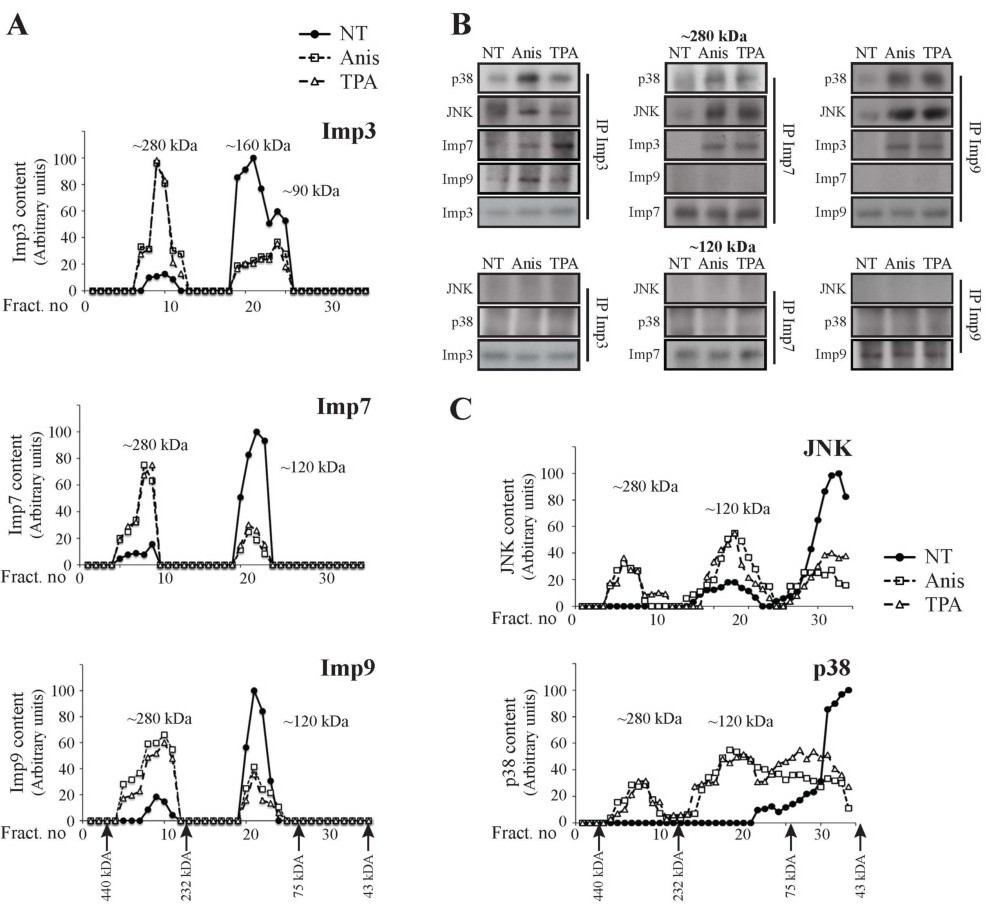
Fig. 10. JNKs and p38s form complexes with dimers of either Imp3/7 or Imp3/9 after stimulation. (A) Gel filtration studies reveal MW shift of Imps 3, 7 and 9 upon stimulation. HeLa cells were serum starved and stimulated with anisomycin, TPA or left untreated (NT). Cell extracts (20 mg) were loaded on a 16/60 superdex 200 sizing column (1 ml/min), and 1ml fractions were collected. The fractions were then analyzed using Western blotting with the indicated Abs. (B) CoIP confirms association of JNKs and p38s with Imps dimers in the ~280 kDa but not the ~120 kDa peaks. Fractions representing the ~280 kDa and ~120 kDa peaks (fractions no. 9 and 22) from each of the Anis, TPA or non- treated stimulated columns, were subjected to CoIP with the indicated Imps Abs. The interacting proteins were detected using Western blotting with the indicated Abs. (C) JNK and p38 MAPKs form complexes upon stimulation. HeLa cells were serum starved and then stimulated with anisomycin, TPA or left untreated (NT). Cell extracts were loaded on a 16/60 superdex 200 sizing column (flow rate 1ml/min), and 1ml fractions were collected. The fractions were then analyzed, using Western blot with the anti JNK/p38 Abs, as indicated.